


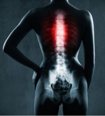
Thoracic osteochondrosis is a degenerative disease of the spine (depletion and destruction of the bone structure of the vertebrae). It starts with impaired posture, the appearance of autonomic symptoms (shortness of breath, weakness, sweating, malaise) and the development of severe pain syndrome. Sternal osteochondrosis is similar to cardiovascular disease, so accurate differential diagnosis is required. Treatment includes a wide range of treatments: medicine, exercise therapy, physical therapy and massage.
Thoracic osteochondrosis is less common than cervical or lumbar spine. This is due to the particularity of the anatomical structure. The thoracic intervertebral disc occupies two thirds of the entire spine, and the diameter is also larger, but smaller than the lumbar spine. This area is strong and low in mobility, and is protected by the rib cage and ribs. The physiological curve points to the back. This leads to increased pressure on the front of the spine. In addition, pathological bone structure formation and growth appear on the vertebral body (osteophytes). Peripheral nerve endings are located between ligaments and muscle tissue, and their tension causes compression as the pain develops.
There are also multi-segmental lesions of the spine with osteochondrosis. At the same time, degeneration of the neck, chest, and waist areas is accompanied by corresponding clinical symptoms.
The clinical symptoms of thoracic osteochondrosis in women and men are roughly the same, and there is no significant difference.
Prevalence
A diagnosis can be made at any age. This disease is common among teenagers with weak musculoskeletal systems and is also the result of their active growth. Due to the significant load on the chest area during pregnancy, pathology is usually developed in pregnant women.
Everyone has a tendency to develop thoracic osteochondrosis. This is due to a person's upright posture, so the spine part bears a lot of load.
classification
Chest pain syndrome is characterized by severe, severe chest pain. The syndrome is related to peripheral nerve damage. Failure is due to the compression of nerves by muscles and ligaments.
Degree of thoracic osteochondrosis:
- The first degree is characterized by no obvious clinical manifestations. The intervertebral disc loses its elasticity and forms a protrusion.
- The second degree is characterized by a further loss of the elasticity of the intervertebral disc and a decrease in height. The possibility of hernia increases. Pain syndrome occurs, and accompanying pain symptoms may occur.
- In the third degree, pain syndromes increase. A herniated disc located between the vertebrae is possible. The severity of symptoms depends on the location of the hernia.
- The fourth degree completely violates the elasticity and loss of intervertebral disc function, and the vertebral bone structure is destroyed. Nervous system diseases are the most obvious.
According to the type of pain symptoms:
- The pathology of the spine proves that the chest pain of the vertebral body is reasonable.
- Non-vertebral chest pain is caused by the formation of internal organs: cardiovascular disease, gastroduodenal reflux, traumatic and inflammatory lesions of the musculoskeletal system.
- Psychogenic chest pain is caused by panic attacks and damage to neurogenic organs.
Causes and risk factors
Without pathological factors, osteochondrosis will not develop. Many reasons or their combination cause the disease to develop in the chest area.
- Sedentary lifestyle. Lack of physical exercise can cause muscle weakness in the back and intervertebral segments. Sedentary immobility and improper workplace organization are another factor leading to sternal osteochondrosis.
- Improper weightlifting and various injuries. Excessive pressure can destroy the function of the spine. In this case, the muscles and intervertebral discs cannot bear the load.
- Acquired lesions and curvature of the spine. In the context of these pathologies, the work of the spine is disturbed, and the possibility of osteochondrosis is increased. If the doctor’s advice is not followed, the damage will increase.
- Lack of essential minerals and vitamins. Due to insufficient calcium concentration in bone tissue, bones become weak and the possibility of damage to the musculoskeletal system increases.
- Pregnancy is a combination of major factors: increased spinal load and lack of minerals and vitamins.
important!Genetic predisposition plays an important role. If the pathological changes of the musculoskeletal system are observed along the relevant line, then you should pay attention to your health and prevent pathological changes. An effective system of preventive measures can prevent large-scale destruction of bone tissue.
Who is at risk
Usually, the factors that form the degenerative changes of the spine are combined.
- Decreased immune status is associated with increased susceptibility to infection, and can enhance the clinical manifestations of osteochondrosis caused by muscle inflammation.
- Pressure effects that can cause psychogenic chest pain. This is due to the increased release of catecholamines which leads to increased pain.
- Damage to the nervous system caused by non-infectious and infectious diseases.
- The body is overloaded.
- Does not conform to ergonomic principles (weight bearing).
- Spinal injuries of various reasons.
- Muscle cramps.
- Osteoporosis of the musculoskeletal system.
symptom
The main symptoms of thoracic osteochondrosis
- The intercostal space produces a burning sensation.
- Paroxysmal and persistent pain in the chest, mainly tingling.
- For chest pain, the pain syndrome manifests as tingling, contraction, and pain.
- Low back pain.
- Pain on one side of the torso.
- During the exercise, you will notice the tightening of the vertebrae.
- Pain symptoms increase significantly with exercise, deep breathing, coughing, and sneezing. This is the main difference between sternal osteochondrosis and angina.
- The affected area is palpable, that is, they can be felt and located along the affected nerve.
- Numbness of the skin along the intercostal space.
- When exposed to low temperatures or in an uncomfortable position for a long time, the patient's condition will worsen.
Various pain syndromes of thoracic osteochondrosis:
- Lower neck lesions. There is soreness in the upper chest, which can radiate to the neck, arms and the left half of the body.
- Upper thoracic spine injury. Pain is pain in nature and affects the central part of the chest. Often combined with pain in the scapula area.
- Failure of the scapular region. Pain symptoms are characterized by cutting, pain, and tingling. Has an offensive appearance, whether long or short. It occupies the lateral area and is also concentrated in the scapula area.
- Pain in the anterior chest wall, varying in duration. They appear between the thoracic and frontal axillary lines.
In addition to the main symptoms, there are two types of pain syndromes in thoracic osteochondrosis:
- Dorsago-Intense but short-term pain at the location of the affected intervertebral disc. Normal breathing disorder.
- Back pain-mild but prolonged pain in the affected disc area.
Spinal chest painIt is related to damage to the musculoskeletal system, usually accompanied by severe pain and instability of the thoracic vertebrae (their mobility increases). The failure is manifested in the violation of the mobility of the thoracic spine, the pain of suture and cutting of the intercostal space.
Vertebral chest painCan cause the following symptoms:
- Radical (pain symptoms);
- Violation of the innervation of the chest belt (visceral manifestations: many patients have tingling pain symptoms in the digestive tract or cardiovascular system);
- Nerve root syndrome (intercostal pain) with vegetative signs.
When diagnosing a problem, you need to distinguish the symptoms from cardiovascular disease and myalgia. Ischemic causes of heart damage are characterized by the regularity that occurs during periods of physical or mental-emotional stress and the relief of attacks by taking nitrates.
Psychogenic episodes of chest pain are accompanied by the appearance of panic, anxiety, suffocation and mental disorders. It turns out that this disease is the result of psychological stability problems.
The clinical symptoms of osteochondrosis are divided into two main parts:
- Neurological symptoms:
- For thoracic osteochondrosis, numbness and tingling can occur in the upper limbs and intercostal space, and spread to the front surface of the chest.
- The latissimus dorsi and pectoralis muscles are always in a state of tension.
- There is a high degree of emotional instability, often tears and irritability.
- In rare cases, the disease manifests as obvious intercostal neuralgia.
- Various pain sensations:
- Back: Severe pain in the thoracic spine, and sometimes difficulty breathing. The movement of the cervical and thoracic spine is limited. When sitting in a distorted position, it manifests or deteriorates.
- Back pain: It takes two to three weeks for the formation of pain symptoms. Therefore, it has no clinical manifestations for the patient at first. Slight discomfort in the chest. Turning your body to the sides and breathing deeply will exacerbate the pain. With the final stabilization of the pathological process, a persistent pain syndrome is formed.
- Intercostal neuralgia: belt pain radiating along the intercostal space. When you breathe quickly, there will be a tingling sensation in the heart area. Therefore, pathology is often confused with damage to the cardiovascular system.
- With the development of reflex angina pectoris, lesions appear at the level of the Th1 segment, forming the heart or pseudocoronary syndrome. The difference with organ damage in the cardiovascular system is pain when bending or rotating the spine. They will aggravate with prolonged periods of forced posture. Pain when palpating the spinous process of the thoracic spine.
- Radical syndrome: soreness in the intercostal space (Erb point).
- Visceral syndrome: Abdominal organ dysfunction, disease at the level of the V-XII thoracic spine. It manifests as a pain in the waistband, a heaviness on the right side of the flanks, and heartburn.
The clinical symptoms depend on the level of thoracic spine lesions:
* Failure of the neural process in sternal osteochondrosis occurs when osteophytes appear-bone growth on the vertebrae. This is due to the rate of destruction. Therefore, the following symptoms are not part of the disease.
- Nerve mutations at Th2 and Th3 levels. With the onset of arrhythmia and coronary heart disease, the cardiovascular system will be damaged. Therefore, chronic pain symptoms in chest pain can cause organ dysfunction in the cardiovascular system.
- Defeated at Th4-Th5 level. Organs with damaged nerve fibers: pleurisy and bronchitis, pneumonia, bronchial asthma.
- Th5-Th6: The bile duct and gallbladder are affected. The absorption of fat in the body is reduced.
- Th6-Th7: Affect the liver and solar plexus area. Hepatobiliary function is impaired.
- Th7-Th8: The stomach is affected. Indications: Duodenal and gastric ulcer disease, dyspepsia, gastritis.
- Th8-Th9: Changes in the function of the duodenum and pancreas. Manifestations: duodenitis, pancreatitis and loose stools.
- Th9-Th10: damage the nerve cells of the internal organs (spleen and diaphragm). Hiccups and breathing problems will occur.
- Th10-Th11: The adrenal glands are affected. The activity of the immune system decreases and allergies appear.
- Th11-Th12: Impaired renal function, leading to the formation of pyelonephritis and urolithiasis.
- Th12-L1 (the level of the first lumbar spine). The kidneys and ureters are damaged. This can cause difficulty urinating-problems with urination.
Diagnosis of thoracic osteochondrosis
If you suspect osteochondrosis, you can contact a therapist or neurologist.
Check the patient and record all clinical data. During the formation of stages 2-3, the skeleton has undergone significant deformation. The patient’s complete medical history should be collected in order to accurately determine or rule out the factors leading to the formation of thoracic osteochondrosis.
The first method of diagnosis is radiography. According to the clinical history data and the need for differential diagnosis, further research should be carried out.
Any doctor can conduct a preliminary examination of the patient. The main thing is a competent and fully collected clinical history. This will enable you to accurately determine the cause of the disease and choose treatment options. Therapists, neurologists, and rheumatologists are all involved in the treatment of sternal osteochondrosis. If trauma is caused to the spine area, you need to consult a traumatologist.
- X-ray examination of the chest in two projections. Allows you to determine the presence and size of osteophytes, determine the contour and height of the intervertebral disc, and determine the change in the shape of the intervertebral disc.
- Discography can examine the structure of the nucleus pulposus by using contrast.
- Computed tomography is used to show nerve fibers, muscles, ligaments and joints.
- Electromyography can be used for differential diagnosis with neurological diseases.
- The endoscopic diagnosis method can be prescribed to examine the circulation and digestive organs.
- Perform an electrocardiogram to determine the cause of cardiovascular disease.
- EEG-to establish the pathology of the nervous system.
Differential diagnosis
Sternal osteochondrosis should be distinguished from many diseases.
- Abnormal spine formation, trauma, tumor, inflammation. There are many options for these pathologies. For example, additional congenital processes, vertebrae displacement or fusion (spondylolisthesis), osteomyelitis, ankylosing spondylitis, etc.
- Damage to the musculoskeletal system (different lower limb length, muscle cramps, muscle inflammation, etc. ).
- It has nothing to do with damage to the musculoskeletal system, but is similar to the symptoms of internal organ diseases. Especially pancreatitis, adnexitis, gastric ulcer, coronary heart disease, angina pectoris, and pleurisy.
- Neurosis-like disease, accompanied by migratory pain, increased fatigue, irritability, and mood swings.
Thoracic osteochondrosis and ischemic heart disease
An effective differential diagnosis of the most similar pathology is extremely important. There are many differences between the pain caused by vertebral chest pain and coronary heart disease (IHD), which makes accurate diagnosis possible.
The nature of pain: When suffering from coronary artery disease, they have the characteristics of burning and contraction, and accompanied by the fear of death.
According to the duration of pain:
- IHD: Short-term, onset within a few minutes.
- Thoracic osteochondrosis is characterized by subsided or persistent pain. In some cases, they will not go away during the day.
Position changes:
- For ischemic heart disease, the intensity and intensity of pain do not change with physical activity.
- For chest pain, even relatively mild exercise can cause the pain to increase or new attacks occur.
Response to physical activity:
- For ischemic heart disease, pain occurs during physical exertion and stops at rest.
- On the contrary, chest pain will diminish, but it will not stop.
Cupping medication:
- For ischemic attacks, taking nitrates is easy to relieve pain.
- The use of analgesics can relieve chest pain.
The influence of physical therapy factors and manual therapy:
- For ischemic heart disease, it will provide instability and slight improvement.
- For osteochondrosis, the patient’s condition has a markedly positive dynamic.
Treatment of chest osteochondrosis
Osteochondrosis is treated by a neurologist.
In order to organize effective treatment, the prerequisites for etiology need to be established first. Determining the cause of the pathology allows you to choose the right treatment.
Taking into account all the functional characteristics of the body, the preparation of bone tissue regeneration is selected. It is recommended to clarify the concentration of collagen and elastic fibers in the body in advance. When choosing a treatment plan, the individual characteristics of the organism will be considered.
Standard treatment plan
Non-steroidal anti-inflammatory drugs help relieve chest pain caused by inflammation. This increases the amount of activity in the chest and the range of motion of the thoracic spine.
Drugs that affect the production of interleukins. They can stop the inflammatory cascade and normalize the balance of enzymes that lead to the destruction of nerve myelin.
Antispasmodics are also used.
B vitamins help prevent inflammation of the affected nerves.
Preparations that preserve the concentration of collagen and elastic fibers allow you to keep the fluid in the intervertebral discs. This increases tissue elasticity and prevents further degradation.
Hormonal (steroid) drugs. They have a powerful anti-inflammatory effect, but are only used for acute chest pain because they have a negative effect on the entire body.
Diuretics in the acute phase of the disease help relieve swelling of nerve endings. Potassium-sparing diuretics are preferred.
Anti-inflammatory ointments and gels. When rubbing the affected area on the back, the local inflammatory process is reduced and the highly active pain symptoms disappear.
massage
The therapeutic effect of massage is to relieve the spasm of thoracic spine muscle bundles and normalize local blood circulation.
The effect of massage technique:
- Remove excessive muscle tone;
- Strengthen the body structure of the intervertebral disc.
The use of massage techniques is combined with visits to chiropractors and a regular exercise therapy system.
physiotherapy
acupuncture. Eliminate or reduce muscle cramps and relieve pain symptoms.
Manipulative therapy. Allows you to restore the systemic circulation to a normal state in the intercostal space. This regulates the nutrient supply of the tissues, improves their nutrition and stimulates the oxygenation of the blood.
Nutrition of thoracic osteochondrosis
Observing certain nutritional principles can allow you to achieve the maximum therapeutic effect.
- Recommend foods rich in vitamins A, B, C and E (vegetables, nuts, grains).
- Omega-3. 6 fatty acids found in fish.
- The cartilage tissue regeneration stimulator in the form of food additives can maintain the strength of the tissue and the elasticity of the tissue structure.
complication
When determining the diagnosis of thoracic osteochondrosis, a series of possible organ diseases that may occur over time should be considered.
- Damage to the cardiovascular system: Persistent pain syndrome leads to unstable myocardial ion exchange, which is a prerequisite for coronary heart disease.
- Abdominal organ dysfunction: stomach, duodenum, pancreas. This is due to the excessive secretion of adrenaline in persistent pain syndrome, leading to increased secretion of VIP (vasogut peptide).
- The cause of gallbladder dyskinesia is the increase in bile stone formation in the context of a chronic inflammatory process.
Regularly adhere to the principle of treatment, exercise the treatment system, maintain posture and eliminate risk factors, and the course of the disease will be reduced to subsidence. If the pathology does not develop further and the disease does not actively manifest itself, the prognosis is considered good.
prevention
- Eliminate fatigue, treatment exercises. Choose resistance exercises, vertical loads with displacement, and spine extension.
- When driving a car for a long time, choose a special exercise to relax the muscle skeleton.
- Suction the muscles of the thoracic spine. When independent training is not possible, there is the use of exercise therapy complexes and muscle stimulation.
- Organization of the workplace: The backrest of the work chair should provide support for the spine. In order to keep the load on the spine from increasing, you should warm up by stretching or walking every 30 minutes. This is because sitting posture puts more pressure on the spine.
- The correct position of the spine at night: buy orthopedic accessories for sleep. Due to the violation of the physiological curve of the spine, a completely rigid surface is unreasonable.
- Observe the principles of ergonomics: do not lift heavy objects that may harm the spine.
- Formation of the correct posture.
- Optimize blood circulation and lymphatic flow through the stretch mark system or the use of special procedures (compression therapy).